Imaging

The old Chinese proverb advising "A picture is worth a thousand words" is no more amply demonstrated than in the visual information found in magnified images. The crucial detail revealed by extreme close-up or macro photographic and microscopic imaging of failed or unidentified materials displays a whole new realm of information for investigators seeking to explain probable root cause failures in processes or materials.

However, because of the unfamiliar or "alien" nature of the visual content of magnified images created either by optical magnification or through manipulation of the properties of light with techniques such as polarization or phase contrast, the relevant data establishing the scale of size or the extent of light alteration needs to be displayed directly on the image being viewed. The presence of the degree of magnification or the cause for the unusual visual appearance of the image, actually being part of the field of view, serves to normalize or restore the observers visual perception.

Current technology using large scale integrated circuitry, impressive computing power and image processing software makes possible the real time preparation and integration of both macro and microscopic images with the physical data describing their creation.

Charge coupled devices capable of five mega pixel, full colour resolution can be coupled to cabled or wireless desk top or notebook computing systems with USB technology to capture real time video or still images from cameras on low or high powered microscopy systems using plain or highly manipulated light sources for illumination.

Civil forensic science investigations usually do not allow for destructive materials testing so as various experts can independently examine the evidence at hand. Physics, generally speaking, teaches that high powers of magnification with visible light are obtained at short viewing distances. Fortunately current digital viewing systems permit long USB cables to separate the optical magnification system from the visual display and recording functions. Thus it is often possible to remove the optical viewing components from a desk top mounting stand and with some imaginative arrangements of clamps and supporting structure, produce magnified images of large objects such as failed pieces of furniture or massive vehicle components.

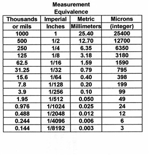
  Small distances, in the metric system are traditionally measured in microns or 1/1,000,000 of a meter. Small distances in the imperial system are traditionally measured in mils or 1/1,000 of an inch. With the conversion factor of 2.54 centimeters per inch we are able to compile the table of measurement equivalence at the left.

Examination of opaque surfaces or objects that do not transmit light requires the use of special techniques. At low powers of magnification the viewing lenses are usually at a distance far enough from the object being examined so that room light or high intensity lamps can be used to illuminate the surface being examined. For higher powers of magnification the lens must be much closer to the surface being observed and the microscope and lens block the light needed to see the surface. High powers of magnification for opaque materials are obtained by a technique known as vertical illumination in which the viewing lens itself provides the light required to illuminate the subject.

Although microscopy and integrated circuitry have radically altered the time required to capture and edit magnified images and the powers of magnification available are large, if required, the majority of civil forensic science problems are often solved with low to moderate degrees of image magnification.
 

These photos illustrate the crystallized metallic structure of a failed stainless steel on the left and the pair of photos on the right depict two shards from failed plate glasses. The longer shard is from a tempered glass while the short is from regular plate glass. Two photos at different exposures are better able to depict the wide differences in polarization colour intensity. The high degree of surface stress that gives the tempered plate its strength is demonstrated in the intense length wise banding of the fragment, while the weak chaotic un-ordered colours of the short plate glass fragment are the resulting residual stress from the original fracture. All three photos were taken with a hand held digital camera.